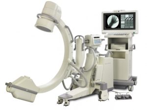
Philips BV Libra

Category:

This is a compact, portable Philips fluoroscopy c-arm that can be used for diagnostics and surgical procedures. The cost-effective, ergonomically-designed and versatile X-ray imaging system delivers high-quality fluoroscopic images while requiring significantly less radiation dose.

Buy / Sell All Models


Philips BV Libra Specifications


X-Ray Tube / Tank Unit
  • Tube type: Fixed anode
  • Nominal focal spot values (IEC 336): 6 IEC and 1.4 IEC
  • Nominal X-ray tube voltage: 110 kV
  • Maximum anode heat content: 5 kJ = 50 kHU
  • Anode cooling capacity: 6 kJ/min. = 30.6 kHU/min.
  • Maximum housing heat content: 840 kJ = 1200 kHU
  • Inherent filtration: 0 Al eq.
  • Additional filtration: 1 mm Al + 0.1 mm Cu

Collimator Unit
  • Iris collimator
    • Type: Circular opening, lead iris leaves
    • Indication: During LIH (and also on image)
    • Shutters
    • Type: 2 independently movable real lead shutters with steel wedge tip
    • Rotation: 360°
    • Indication: During LIH (and also on image)

X-Ray Generatorbe Type:  Fixed Anode
  • Generator type: DC converter, Constant Potential (CP) generator, micro-processor controlled
  • Maximum generator output: 15 kW
  • Maximum X-ray tube voltage: 110 kV
  • Maximum X-ray tube current: 20 mA (30 mA for France)

Continuous Fluoroscopy
  • kV range: 40 to 110 kV
  • mA range for Low Dose Fluoroscopy mode: 10 to 3.00 mA (up to 7.20 mA during Auto High Penetration)
  • mA range for High Definition Fluoroscopy mode: 24 to 7.20 mA

Pulsed Fluoroscopy
  • kV range: 40 to 110 kV
  • mA peak range: 10 to 3.00 mA (up to 7.20 mA for High Penetration)
  • Pulse width: 40 ms
  • Pulse rates: 12.5 pulses/second

Intermittent Fluoroscopy
  • Pulse time: 240 ms
  • Pulse rate: 1 PPS (pulse per second)
  • kV range: 40 kV – 110 kV

Radiography
  • kV range: 40 – 105 kV
  • mA range: 20 mA fixed (30 mA for France)
  • mA range (R’10 series from ISO 497): 0 to 80 mAs

Detection (II/TV)
  • Image intensifier type: Single mode 6” HC, Triple mode 9” HRC
  • Nominal image intensifier formats 6”: 15 cm
    • 9”: 23, 17, and 14 cm (9”, 7” and 5”)
  • Entrance screen = Input screen: Cesium Iodine
  • Detection Quantum Efficiency (DQE), typical [%] according to IEC 1262-5: 6”: 60, 9”: 62
  • Grid type: Circular, carbon fiber; 60 lines/cm
    • Ratio = 1:10 at FFD = 100 cm
  • TV camera type: Interline transfer CCD; high resolution
  • Lens type: Anamorphic lens
  • Image rotation by camera: 400° : +/- 200°
  • Rotation position indication: Pre- indication on image, during LIH
  • Video standard CCIR: 625 lines; 2:1 interlaced
  • Image reversal: Included
  • Automatic anatomical measuring field: Included with ‘BodySmart’

TV Monitors
  • Type: Standard high quality monitors High resolution, high contrast, 17″screen size, 75 Hz non-interlaced scan frequency
  • Type: High end monitors High resolution, high contrast , extra high brightness, 17″screen size, 75 Hz non-interlaced scan frequency, Triple Gun Technology
  • Type: Standard color LCD monitors Extra high resolution, high contrast, 18″screen size, TFT technology, resolution 1280×1024 (hxv), 250 cd/m2
  • Type: High brightness color LCD monitors. Extra high resolution, high contrast, extra high brightness, 18″screen size, TFT technology, resolution 1280×1024 (hxv), 500 cd/m2 . Both standard and high brightness color LCD monitors can be optionally mounted on a flexible arm for optimal image presentation.


Image Storage and Processing
  • Digital Image Processor type: Dedicated 12 bit video pipeline processor
  • Display image matrix size: 1008 x 560 x 8
  • Image storage capacity and max. storage rate: 1,000 images (max.5 frames/second) 500 images (max.3 frames/second 50 images (max. 3 frames/second) 16 image, RAM memory (standard)
  • Patient data handling: Multi-patient database
  • Image processing: Edge enhancement (real-time),Windowing (real-time), Recursive noise reduction, Movement detection, Mosaic, Replay
  • Processing options: Subtraction, Remasking, Edge enhancement (post),Windowing (post),Annotation, Zoom, Measurement, Electronic shuttering

Geometry
  • Longitudinal movement: 200 mm
  • Swivel range: ± 10°
  • Vertical movement: 490 mm (+410 mm/-80 mm) Motorized
  • Rotation: ± 180°, with safety stop at ± 135°
  • Angulation (orbital movement): +90°, -25°
  • Source to image distance (SID): 995 mm
  • Free space within C-arc: 780 mm
  • C-arc: depth 630 mm
  • Brakes for all movements: Included, manual
  • Steering: Rear wheel
  • Parallel movement: Via rear wheel control
  • Cable deflectors: Included
  • C-arm stand weight: 9”: 260 kg; 6”: 260 kg
  • C-arm stand length 9”: 1815 mm; 6”: 1785 mm
  • C-arm stand width: 890 mm
  • C-arm stand height: 1680 mm
  • Mobile View Station depth: 760 mm
  • Mobile View Station width: 930 mm
  • Mobile View Station height: 1850 mm

Power Supply
  • Input voltage: 110-240 V +/- 10%
  • Frequency: 50/60 Hz

Options
  • Laser alignment tool: Yes
  • Laser aiming device: Yes (9” only)
  • Video paper/transparency printer: Yes
  • Video DVD Recorder: Yes
  • ViewForum Surgical Workstation: Yes (supports DICOM Query/Retrieve, DVD DICOM Store, Procedure Reporting Package and MIP/MPR)
  • Standard DICOM package: Yes (supports DICOM print, DICOM store)
  • Advanced DICOM package: Yes (incl. MWL, MPPS, SC)
  • Sterile covers: Yes
  • Detachable cassette holder: Yes
  • High End monitors: (triple gun) Yes
  • 18″ Color LCD monitors Yes


Bluestone Diagnostics - Product Form

  • This field is for validation purposes and should be left unchanged.