|
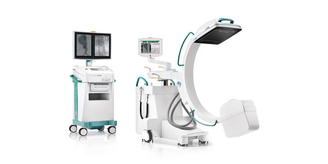
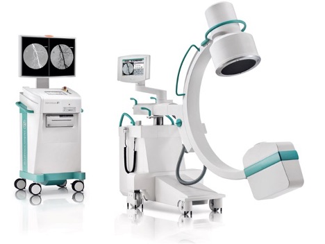
GE OEC Elite Specifications
|
|
|
Image Acquisition |
-
9-Inch Image Intensifier
- Tri-mode 9”/6”/4.5” image intensifier
- Minimum central resolution (at monitor):
- 9”: 2.2 lp/mm
- 6”: 3.0 lp/mm
- 5”: 3.5 lp/mm
- DQE: 65% (typical)
- Removable grid
|
|
Image Processing |
- 1 k x 1 k x 16 bit: image intensifier
|
|
Generator |
- 60 kHz high frequency
- 15 kW power
- Up to 120 kVp
- Continuous high level fluoroscopy (HLF) up to 20 mA
- Pulsed HLF up to 40 mA
- Digital spot up to 75 mA
- Full power from a standard wall outlet
- Patented battery buffered design
|
|
X-Ray Tube |
- Rotating anode X-ray tube
- 3 mm and 0.6 mm nominal focal spots
- Anode heat capacity: 300,000 HU
- Anode cooling rate: 85,000 HU/min
- Housing heat capacity: 1,600,000 HU/min
- Housing cooling rate:
- 21 cm CFD – 22,500 HU/min
- 31 cm CFD – 31,000 HU/min
- 9” I.I. – 22,500 HU/min
- 12” I.I. – 22,500 HU/min
|
|
Mainframe |
- System Height: 69.7” (177 cm)
- System Width: 33.5” (85.1 cm)
- System Length: 77.8” (197.6 cm)
- Weight: 610 lbs. (277 kg)
|
|
C-Arm |
- SID: 39.4” (100.1 cm)
- Free space in arc: 31” (78.7 cm)
- Depth in arc: 26” (66 cm)
- Orbital Rotation: 115 ° (90° Underscan / 25° Overscan)
- Lateral Rotation: 360° (+180° / -180°)
- Wig/wag: 20°
- Horizontal travel: 8” (20.3 cm)
- Vertical Travel: 18” (45.7 cm)
|
|
Workstation |
- Height: 67.5” (171.5 cm)
- Width: 34.1” (86.7 cm)
- Depth: 25.8” (65.5 cm)
- Weight: 428 lbs (194 kg)
|
|
Digital Image Orientation |
- Digitally adjusts image display for live and last image hold
- Automatic image update preserves image orientation settings applied during live and last image hold for subsequent images
- Image rotation
- Live and last image hold rotated in real-time
- On-screen display of rotation degrees
- Image reversal (left-to-right)
- Image invert (top-to-bottom)
- On-screen orientation indicator (real-time feedback without fluoroscopy)
|
|
PreView Collimator |
- On-screen collimator position indication
- PreView iris collimator
- PreView Tungsten rotatable double leaf collimator
- Adjust collimators without X-ray exposure
|
|
|
|
|
Fluoroscopy Mode |
- kVp range: 40 – 120
- mA range: 0.2 – 10 normal mode 0.2 – 20 HLF
- Auto and manual fluoroscopy modes
|
|
Pulsed Fluoroscopy Mode |
- kVp range: 40 – 120
- mA range: 0.2 – 10 normal mode 0.2 – 40 HLF
- Pulse rate: 8 pps
- Pulse width: 25 ms
- Auto and manual pulsed fluoroscopy modes
|
|
Digital Spot Mode |
- kVp range: 40 – 120
- mA range: Up to 75
- Automatic exposure termination and automatic image save
|
|
TechView Monitor |
-
*Optional on image Intensifier modes
- 4” (26.4cm) display, LCD flat panel monitor mounted on mainframe
- 270° side/side rotation
- 30° up/ 5° down tilt
- Horizontal viewing angle 80°, Vertical viewing angle 70°
- 800 x 600 resolution monitor
|
|
AutoTrak |
- Automatically seeks the subject anatomy anywhere within the imaging field
- Selects the optimum imaging technique by varying mA, kVp, and gain
- Automatically adjusts to anatomical size and location
- Provides uniform image quality throughout the entire image
|
|
Image I.Q. |
- Smart Window – Dynamically senses the collimator position and automatically adjusts image brightness and contrast
- Smart Metal – Adjusts brightness and contrast automatically and allows the user to adjust sensitivity levels for optimum image quality even when metal is introduced into the field
- Tungsten Collimator – Denser collimator limits X-ray exposure area, reduces scatter radiation, and improves image detail
|
|
Connectivity |
- Wired Ethernet
- USB data transfer
- DVI-I output
- Integrated film/paper printer (optional)*
- Thermal printers
|
|
Video Monitor |
- 27 in (69 cm) LCD TFT color display
- Anti-glare
- 450 cd/m2 maximum brightness
- Touch screen system control
- 2560 x 1440 high resolution display
- Integrated PIP window to display color
- DVI-D input
|
|
User Interface |
- Touchscreen control simplifies operation
- Automated system operation requires minimum operator interface
- Multi-functional controls with a footswitch or hand-held control
- Physical keyboard with integrated touchpad with a sealed silicone design for dust-free, contaminant-free and water-resistant use
- Physical image control keypad
- On-screen virtual keyboard and image control keypad
- Multi-purpose image directory to retrieve and review images, copy image(s), or manual delete image(s)
- Integrated DICOM interface
- Radiation dose structured report (RDSR)
- X-ray dose summary
- Room-in-use indicator interface
- SmartConnect allows workstation to operate independently of C-arm and connect/ disconnect C-arm when needed
|