|
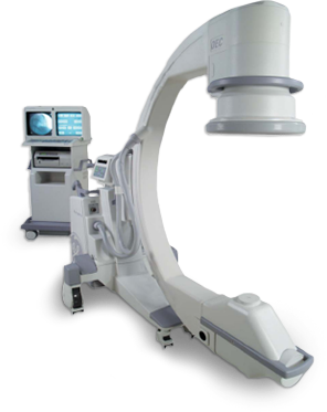
GE OEC Brivo Plus Specifications |
|
|
C-Arm |
- SID: 4″ (100 cm)
- Free Space in Arc: 7″ 78 cm)
- Depth in Arc: 26″ (66 cm)
- Orbital Rotation: 120° (90° underscan / 30° overscan)
- Lateral Rotation: 410° (+205° / -12.5°)
- Lateral Height: 2″ (102 cm)
- Wig / Wag: 20° (+12.5° / – 12.5°)
- Horizontal Travel: 8″ (20 cm)
- Vertical Travel: 5″ (44.5 cm)
|
|
X-Ray System – Generator |
- 40kHz high frequency
- 2kW monoblock
- Housing heat capacity: 900,000HU
- Housing Cooling Rate: 12,500HU/min
- Up to 110kVp
- Up to 20mA for radiographic film exposure
- Continuous high-level fluoro (HLF) up to 12mA
- Digital spot up to 16mA
|
|
X-Ray Tube |
- Stationary anode X-ray tube
- Dual focal spot:
- Small Focal Spot: 0.6mm x 1.4mm
- Large Focal Spot: 1.4mm x 1.4mm
- Tube assembly total filtration: 35mm Al
- Anode Heat Capacity: 76,000HU
- Maximum anode cooling rate: 37,000HU/min
- On-screen tube heat capacity indicator
|
|
PreView Collimator |
- On-screen collimator position indication
- PreView iris collimator
- PreView tungsten rotatable double leaf collimator
- Adjusts collimators without X-ray exposure
- Can reduce X-ray dose to patient and operator
|
|
Low Dose Pulsed Fluoro |
- kVp range: 40 -110
- mA range: 1 – 2
- Pulse rate: 1, 2, 4, 8 PPS
- Auto and manual fluoro modes
|
|
High Level Pulsed Fluoro |
- kVp range: 40 -110
- mA range: up to 12
- Pulse rate: 1, 2, 4, 8 PPS
- Auto and manual fluoro modes
|
|
Digital Spot Mode |
- kVp range: 40 – 110
- Up to 10mA for 100-120V system
- Automatic exposure termination and automatic image save
|
|
Radiographic Mode |
- mA range: 20 (10 for 100-120V system)
- mAs range: up to 80 (40 for 100-120V system)
- Computer controlled exposure time
- Film cassette holder: 10” x 12” (25.4cm x 30.5cm)
|
|
Video Imaging System
Precise-Imaging features
|
- AutoTrak
- Automatically seeks the subject anatomy anywhere within the imaging field and selects the optimum imaging technique
- Automatically adjusts to anatomical size and location
- Provides uniform image quality throughout the entire image
- Simplifies operation
- Smart Window
- Dynamically senses the collimator position and automatically adjusts brightness and contrast to produce high image quality
- Smart Metal
- Automatically detects the metal in the field and optimizes image quality
- Allows the user to adjust automatic brightness and contrast sensitivity levels to metal
- Provides optimum image quality even when metal is introduced to the field
|
|
|
|
|
9” Image Intensifier |
- Tri-mode 9”/6”/4.5” (23cm/15cm/11cm) image intensifier, The central resolution (typical value at image intensifier):
- 9” (23cm): 52 lp/cm
- 6” (15cm): 58 lp/cm
- 5” (11cm): 68 lp/cm
- Minimum central resolution (at monitor):
- 9” (23cm): 2.2 lp/mm
- 6” (15cm): 3.0 lp/mm
- 4.5” (11cm): 3.5 lp/mm
- DQE: 65% (typical)
|
|
Tungsten Collimator |
- Denser collimator limits X-ray exposure area
- Reduces scatter radiation
- Improves image detail
|
|
Tungsten Collimator |
- Denser collimator limits X-ray exposure area
- Reduces scatter radiation
- Improves image detail
|
|
Carbon Fiber Grid |
- 60L/cm 10:1 anti-scatter grid
- Reduces scatter radiation effect
- Improves image detail
|
|
Video Camera |
- High-resolution 1k x 1k CCD camera
- Full digital interface
- Variable gain control
|
|
Video Display |
-
- Dual medical 19” (48cm) monochrome LCD with anti-glare panel.
-
- Tilt motion: 7 degree up / 10 degree down
- Swivel motion: 180 degree
- Viewing angle: 170 degree horizontal and vertical
- Maximum brightness: 1000 Cd/m2
- Maximum contrast: 900:1
- 1280 x 1024 high resolution
- Digital Visual Input (DVI) interface for external monitors
|
|
Image Processing |
|
|
Brilliant-Dose Management Platform |
- High-resolution 1k x 1k CCD camera
- Full digital interface
- Variable gain control
|
|
Additional Features |
Other Image Storage Options
- CD/DVD writer (Optional)
- USB memory stick port
- Integrated DICOM storage
Hardcopy Options
- Integrated film/paper printer (Optional)
- No film developing required
- DICOM printer support
- Multi-format, 1, 2, 4 on 1
- Multi-copy capability
- Radiographic film cassette holder (Optional)
-
Pediatric Application Kit
- Removable grid (Optional)
- Additional filter (Optional)
- Integrated Uninterruptible Power
- Patient Image data protection
- Orderly shutdown
- Electrical
- Input power (60 Hz or 50 Hz)
- 100V/110V/120V @ 20A
- 200V @ 12A
- 220V/230V/240V @ 10A
- Operating Range
- Temperature: 10° to 40°C
- Humidity: 20%-80% non-condensing
|
|
Weight and Dimensions |
- Workstation
- Height: 7″ (167 cm)
- Width: 4″ (90 cm)
- Depth: 25″ (64 cm)
- Weight: 375 lbs (170 kg)
- Monitor Swivel: ±90°
- Monitor Tilt Motion: +7° / -10°
- Mainframe
- System Height: 67.1″ (170.5 cm)
- System Width: 30.7″ (78 cm)
- System Length: 70.3″ (179 cm)
- Weight: 573 lbs (260 kg)
|


